
NIRS Ultra | NIRS Revo | Actus Pro | Actus Lab | HemoX Pro | HemoX Lab
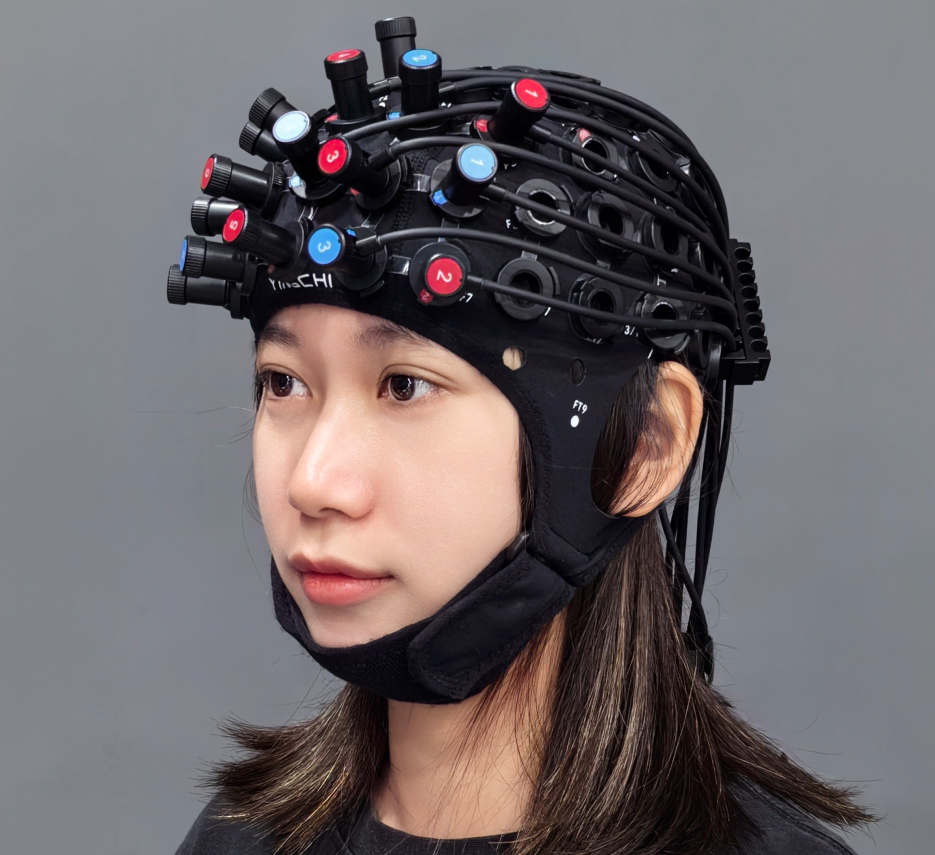
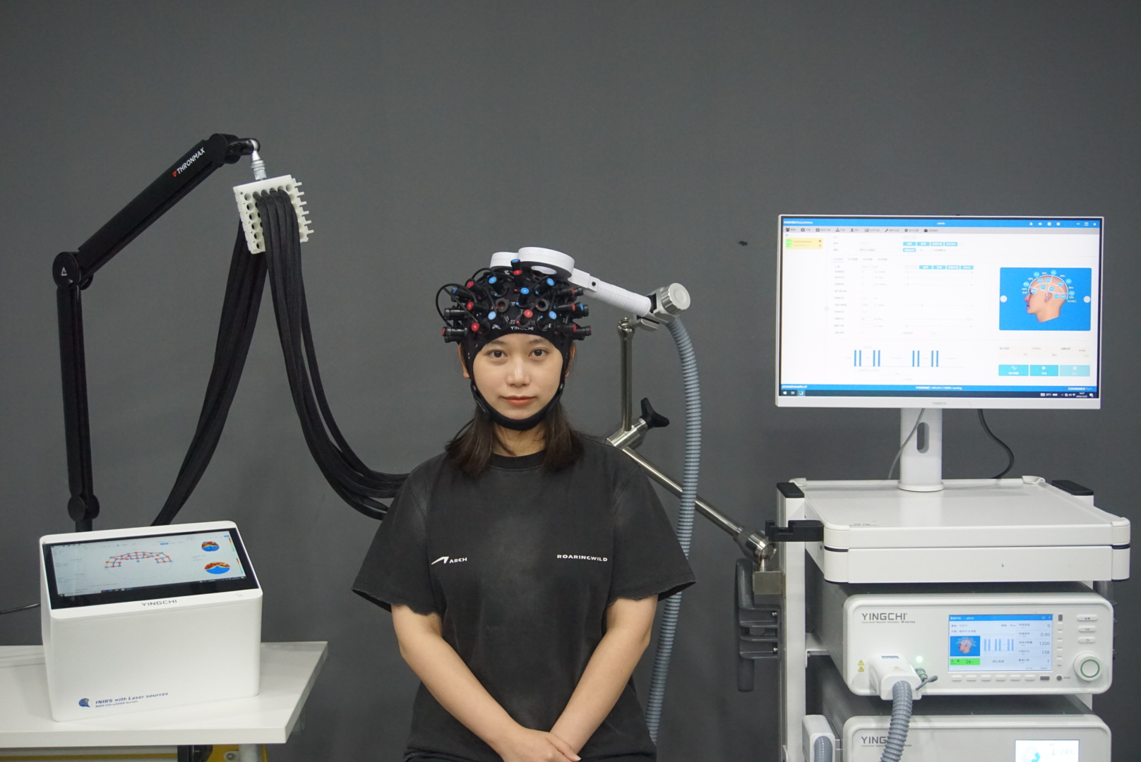
YINGCHI fNIRS — “Your Portable Optical MRI”
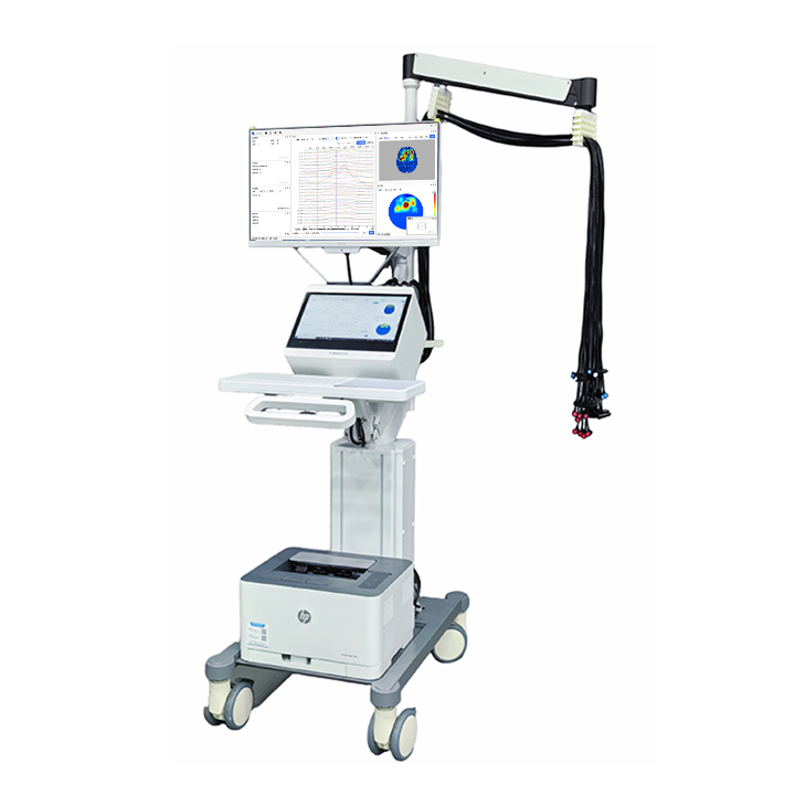
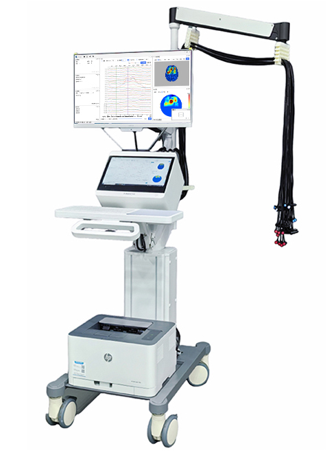
Near-Infrared Functional Brain Imaging System (fNIRS) infers neural activity by measuring hemodynamic changes in the brain. With simple operation, low cost, strong resistance to interference, and high compatibility, it is ideal for brain and cognitive neuroscience research in conventional laboratories, naturalistic environments, as well as for ward-based assessments and clinical brain function evaluation.
Multi-level quality control across hardware and software ensures signal accuracy and reliable mapping of brain activity. >>>
Features of YINGCHI fNIRS
Up to 224 Channels
Configurations support up to 64 sources × 64 detectors with flexible layout for whole-brain coverage.
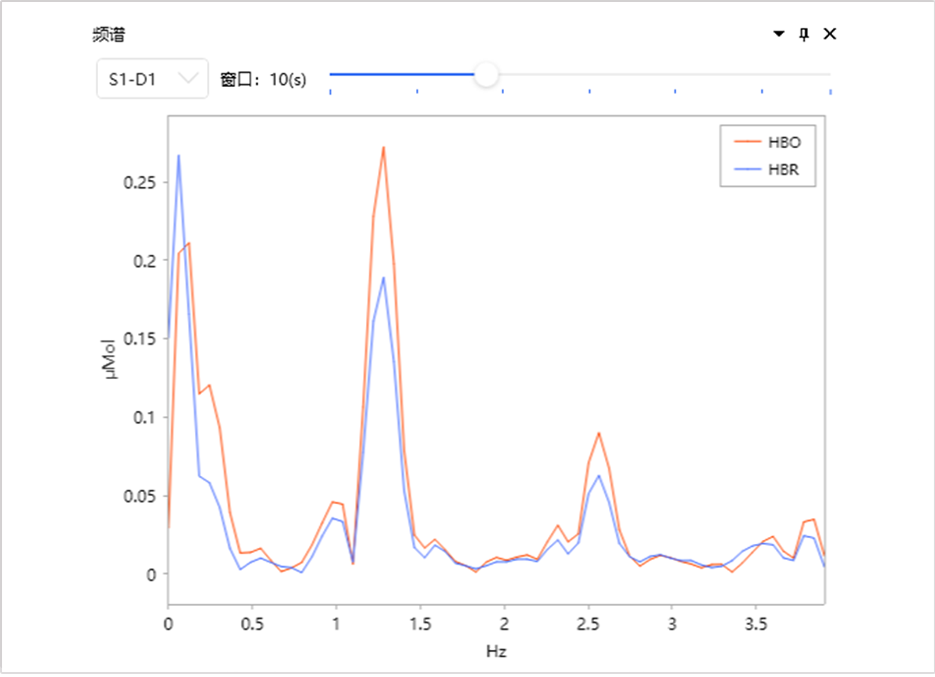
Real-Time Spectral Monitoring
Physiological “fingerprints” of fNIRS—cardiac (~1 Hz) and respiratory (~0.25 Hz) rhythms—are verified in the frequency domain to support robust assessment of signal quality.
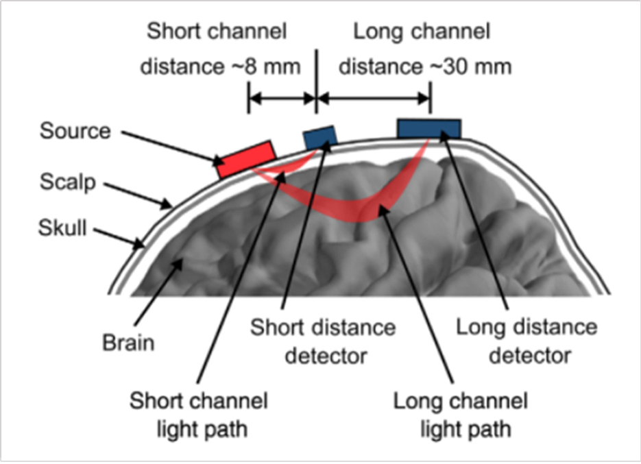
Short-Distance Channels
Removes superficial interference to recover clean cortical signals.
Multimodal Physiological Recording
Respiration, ECG, EDA, and other physiological signals are recorded to interpret brain function in the context of the whole physiological system.
APD Detectors
Avalanche photodiodes enable stable detection of weak hemodynamic signals, ensuring reliable data from deep cortical regions and low-activation tasks.
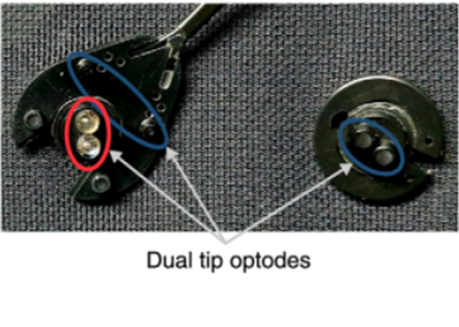
Dual-head Optode Design
Each optode features dual contact points to efficiently separate hair and ensure good scalp contact, reducing preparation time.
Adaptive Three-Stage Optical Probes
Automatically adapt to different hair conditions, ensuring signal quality independent of hair density.
3D Diffuse Optical Tomography (DOT)
Reconstructs three-dimensional hemodynamic changes across different cortical depths.
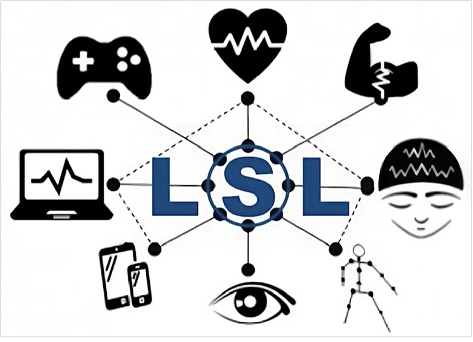
Lab Streaming Layer
Provides high-accuracy temporal alignment for multimodal brain imaging and brain–computer interface applications.
Acquisition & Analysis Software
User-friendly operation with real-time visualization, one-click reporting, and efficient data management.
Scientifically Designed Paradigms
Comes with built-in classical paradigms and also supports parallel testing and adaptive task difficulty, allowing flexible customization for diverse experimental needs.
Open-Source Software Compatibility
Data can be directly accessed and analyzed using third-party open-source platforms.
YINGCHI: Building a Smart, Integrated Brain Science Platform
YINGCHI fNIRS + EEG: Complementary Signals
Synchronized acquisition enables dual-dimensional “electrical–hemodynamic” monitoring, providing complementary insights into brain function.
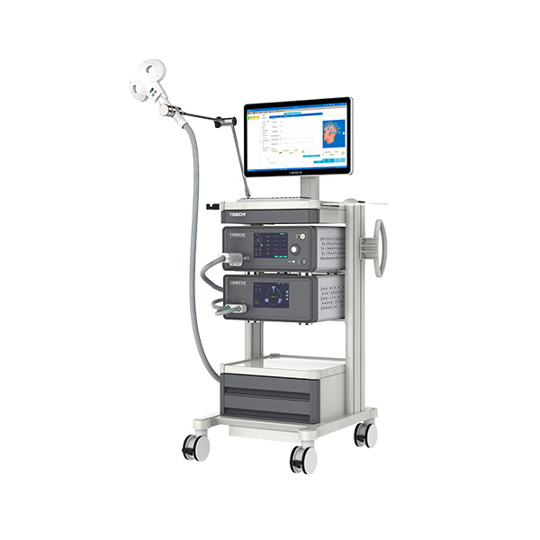
YINGCHI fNIRS + TMS: Causal Research
Noninvasive stimulation techniques combined with real-time hemodynamic recording, forming a “stimulation–response–feedback” closed loop.
YINGCHI fNIRS+psychological/speed–agility training: Integration of Psychology and Behavior
Building a Full-Link “Brain–Mind–Behavior” Evidence Chain.
Technical Applications
DiaBrain–Computer Interface (BCI) and Real-Time Neurofeedbackgnosis
By integrating neurofeedback algorithms with behavioral task modules, the system supports BCI research applications such as motor imagery training, attention modulation, and emotion regulation, demonstrating broad potential in neurorehabilitation and optimization of human–machine interaction.
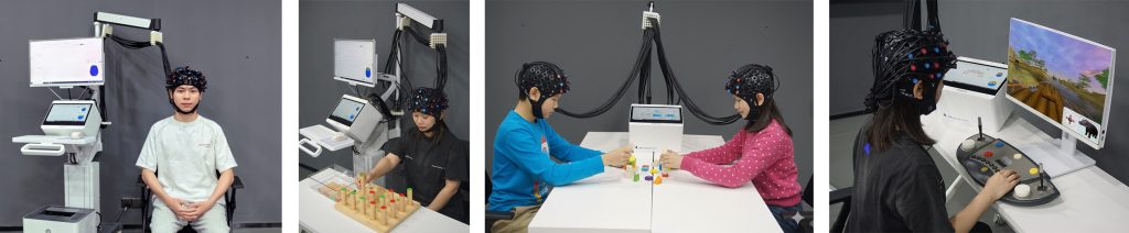
Hyperscanning and Social Interaction Research
YINGCHI fNIRS supports synchronized data acquisition across multiple devices and can be extended to dual- or multi-participant hyperscanning experiments, enabling investigation of brain-to-brain coupling and neural synchronization mechanisms underlying social cooperation and competition.
Cognitive Neuroscience
YINGCHI fNIRS can be used to investigate the neural mechanisms underlying higher-order cognitive processes, including attention, working memory, language processing, and executive functions.
Educational and Developmental Psychology
YINGCHI fNIRS provides a safe and non-invasive method for assessing brain function in children and adolescents, enabling the study of learning, emotion regulation, and neurodevelopmental processes.
Clinical Brain Function Monitoring
YINGCHI fNIRS Provides non-invasive, real-time monitoring of brain activity, enabling dynamic tracking of functional recovery.
Clinical Neurodevelopment and Behavioral Assessment
YINGCHI fNIRS evaluates attention, emotion regulation, and social cognition in children and adolescents, providing support for interventions in neurodevelopmental disorders such as ADHD and ASD.