
Release time :2025-06-25
Source:support@yingchitech.com
Scan:281
On June 10, a team from Fudan University, Department of Neurology at Fudan University Affiliated Pudong Hospital, published new research in Neurotherapeutics (2025 Impact Factor: 6.9): Enhancing working memory in MCI: Modulating alpha-gamma coupling and gamma oscillations via rTMS.
Working memory (WM) is a core cognitive function essential for everyday behavior and complex mental processing. Enhancing its capacity and performance is critically important for improving overall cognitive function. Within WM, visual working memory (VWM) specifically concerns the storage and processing of visual information and is highly dependent on brain oscillations in the alpha (α) and gamma (γ) frequency bands. Alpha waves help suppress irrelevant information and maintain retention, while gamma oscillations support precise memory retrieval, especially under high cognitive load.
Studies have shown that individuals with mild cognitive impairment (MCI) and Alzheimer’s disease (AD) often exhibit impaired occipital gamma activity and reduced VWM capacity, suggesting a close relationship between the two. Cross-frequency coupling (CFC) has increasingly been recognized as a key neural mechanism for integrating information across frequency bands. Specifically, phase-amplitude coupling (PAC) allows alpha oscillations to periodically inhibit or excite neuronal activity, modulating the occurrence of higher-frequency (e.g., gamma) activity.
The alpha–gamma coupling is particularly important for integrating information in short-term memory and plays a crucial role in the frontal–parietal–occipital network, which underlies visual processing and memory tasks. Prior research has found that 10 Hz repetitive transcranial magnetic stimulation (rTMS) applied to the prefrontal cortex can significantly improve cognition in MCI patients. This effect is believed to stem from enhanced alpha–gamma synchrony, which optimizes neural communication and cortical excitability, thereby improving VWM accuracy.
However, the relationship between alpha–gamma coupling and memory load, especially in the MCI population, remains unclear. Furthermore, the dynamic changes in gamma activity across different VWM stages and task demands have not been systematically explored.
This study aims to investigate how alpha–gamma coupling is modulated by varying levels of VWM load and whether rTMS can influence neural oscillatory patterns and memory performance in MCI patients. Using a controlled design, the study compares behavioral and EEG changes in VWM tasks before and after 10 Hz rTMS in both MCI patients and healthy controls. Special focus is placed on the dynamic regulation of occipital gamma activity and prefrontal alpha–gamma coupling, with the goal of providing new theoretical insights and practical avenues for non-pharmacological cognitive enhancement strategies.
This study included a total of 40 participants, 20 MCI patients and 20 healthy controls (HC).
A 64-channel EEG amplifier was used for data collection. During the process, participants performed a Visual Working Memory (VWM) task using an adapted "change detection paradigm" (see Figure 1).
Participants completed 50 practice trials before testing to ensure understanding. Each participant completed 100 trials each of 2T/4T tasks (two blocks of 50 trials), with short breaks between blocks to maintain attention.
Accuracy, response time (RT), and memory capacity (calculated using Cowan's K formula) were recorded. EEG signals were analyzed in three stages: attention (500 ms), encoding (500 ms), and retention (900 ms).
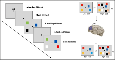
Figure 1: Stimulus Presentation Procedure and Experimental Design of the Visual Change Detection Paradigm ① Attention: Each trial begins with a 200 ms central arrow cue indicating the visual hemifield (left/right) to be memorized. ② Blank: A 300 ms blank interval. ③ Encoding: Presentation of 2 or 4 colored squares for 500 ms (corresponding to 2T/4T load). ④ Retention: A 900 ms blank interval. ⑤ Response: A test image appears, and participants judge whether the color of the squares in the memorized visual hemifield has changed. 2T: 2-target (low memory load); 4T: 4-target (high memory load).
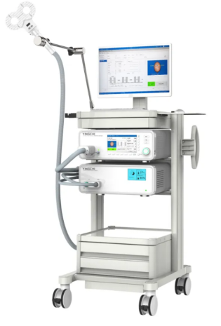
A single rTMS intervention was performed using M-100 transcranial magnetic stimulation device, manufactured by Shenzhen Yingchi Technology, equipped with a figure-8 coil. The specific stimulation parameters are as follows:
| Target | Intensity | Frequency | Train Duration | ITI | Total Pulses | Total Time |
| L-DLPFC | 100%RMT | 10Hz | 10s | 20s | 3000 | 15 min |
▶ Analyzed power spectral density (PSD) in the α band (8–12 Hz) and γ band (30–40 Hz). ▶ To investigate cross-frequency coupling, phase-amplitude coupling (PAC) analysis was performed, quantifying the coupling strength between α phase and γ amplitude using the modulation index (MI). ▶ Evaluated the relationship between right occipital power and VWM performance. ▶ Assessed working memory performance in the VWM task (including accuracy, response time, and memory capacity).
Before the intervention, the MCI group showed significantly lower accuracy and memory capacity in both low memory load (2T) and high memory load (4T) conditions compared to the HC group (P < 0.05, see Figure 2). After a single 10Hz rTMS intervention, the MCI group exhibited a significant improvement in accuracy under the 2T condition, and reaction times were significantly reduced in both 2T and 4T conditions (P < 0.05).
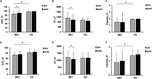
Figure 2: Changes in Working Memory Performance Between MCI and HC Groups. Under low memory load (2T), pre- and post-rTMS intervention comparisons for both groups: (A) overall mean accuracy, (B) response time, and (C) memory capacity. Under high memory load (4T), pre- and post-rTMS intervention comparisons for both groups: (D) overall mean accuracy, (E) response time, and (F) memory capacity. *p < 0.05, pre-intervention vs. post-intervention; #p < 0.05, MCI group vs. HC group. ACC: accuracy; RT: response time.
Under the 2T condition, rTMS induced the following significant changes in EEG activity in the MCI group: during the attention phase, left frontal α-wave power significantly increased; throughout the entire phase, right occipital γ-wave power significantly decreased, with these change trends resembling those in the HC group (see Figure 3A-E). Further Spearman correlation analysis revealed a positive correlation between changes in right occipital γ power and changes in reaction time (P = 0.025, Figure 3F). Before rTMS intervention, phase-amplitude coupling (PAC) between left frontal α and right occipital γ in the MCI group was significantly lower than in the HC group (P = 0.018). Post-intervention, PAC was significantly enhanced (P = 0.034, Figure 3C), suggesting that rTMS can effectively modulate brain oscillations and improve cognitive function. Additionally, baseline PAC levels showed a significant negative correlation with changes in accuracy (P = 0.006, Figure 3G), indicating that lower baseline PAC may predict greater potential benefits from rTMS, holding predictive value.
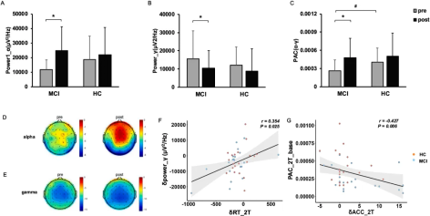
Figure 3: Changes in PSD and PAC in the MCI and HC groups under the 2T condition. (A) Changes in absolute α-wave power in the left frontal lobe before and after TMS stimulation during the working memory attention phase. (B) Changes in absolute γ-wave power in the right occipital lobe before and after TMS stimulation across all phases. (C) Phase-amplitude coupling (PAC) between left frontal α oscillations and right occipital γ oscillations across all phases. (D) Scalp topography of absolute power in the α frequency band. (E) Scalp topography of absolute power in the γ frequency band. (F) Correlation analysis between changes in occipital γ power and changes in reaction time before and after TMS stimulation. (G) Correlation analysis between baseline α-γ PAC and improvement in accuracy after rTMS intervention (post-stimulation vs. pre-stimulation). *p < 0.05: Significant difference before and after stimulation; #p < 0.05: Significant difference between MCI and HC groups; Power 1: Power during the attention phase.
Under the 4T condition, before intervention, the MCI group showed significantly lower left frontal α-wave energy during the attention phase compared to the HC group (P = 0.042). After rTMS intervention, the left frontal α-wave energy in the MCI group significantly increased (P = 0.032, see Figure 4A–B, D–E), while right occipital γ energy showed a decreasing trend. Spearman analysis revealed a positive correlation between changes in right occipital γ-wave energy and improvements in reaction time (P = 0.034, Figure 4F). Although α-γ PAC increased after the intervention, this change did not reach statistical significance (Figure 4C). However, there was a significant positive correlation between post-stimulation PAC and changes in accuracy (P = 0.047, Figure 4G), suggesting that enhancement of α-γ PAC may serve as a neural marker of cognitive improvement.
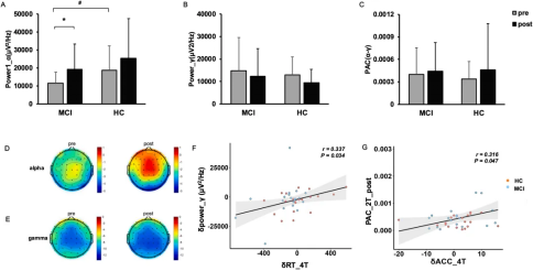
Figure 4: Changes in PSD and PAC in the MCI and HC groups under the 4T condition. (A) Changes in absolute α-wave power in the left frontal lobe before and after TMS stimulation during the working memory attention phase. (B) Changes in absolute γ-wave power in the right occipital lobe before and after TMS stimulation across all phases. (C) Phase-amplitude coupling (PAC) between left frontal α oscillations and right occipital γ oscillations across all phases. (D) Scalp topography of absolute power in the α frequency band. (E) Scalp topography of absolute power in the γ frequency band. (F) Correlation analysis between changes in occipital γ power and changes in reaction time before and after TMS stimulation. (G) Correlation analysis between post-stimulation α-γ PAC and improvement in accuracy after rTMS intervention (post-stimulation vs. pre-stimulation). *p < 0.05 indicates significant differences before and after stimulation; #p < 0.05 indicates significant differences between the MCI and HC groups.
Frontal α activity typically reflects inhibitory control functions, which help suppress irrelevant information to facilitate memory consolidation. Furthermore, functional MRI (fMRI) studies have found that increased α power is associated with decreased BOLD signals in the visual cortex, suggesting a top-down regulatory role during cognitive tasks. These findings highlight the important role of α oscillations in balancing attentional resources and effectively managing cognitive load.
In contrast, γ oscillations—especially in the occipital region—are closely related to attentional processes and the maintenance of sensory information. Changes in oscillatory activity reflect the redistribution of cognitive resources: enhanced frontal α activity helps reduce interference and promotes memory consolidation, while decreased occipital γ activity indicates a reduced demand for visual processing. rTMS stimulation increased frontal α activity and decreased occipital γ activity in individuals with MCI. The former likely reflects improved top-down control, aiding the suppression of distracting information and optimizing cognitive regulation. The latter may indicate a shift of attention from external sensory input to internal cognitive processes, reducing sensory interference and unnecessary high-frequency neural activity, thereby enhancing the ability to focus on tasks and accelerating information processing. This redistribution of cognitive resources may underlie the neural mechanism by which cognitive function improves in MCI patients.
This study also highlights the impact of memory load on phase-amplitude coupling (PAC) between frontal α and occipital γ oscillations. Under low load conditions, due to lower cognitive demands, the neural system has greater regulatory flexibility, allowing PAC to better reflect the efficiency of resource allocation and cross-regional coordination, showing significant changes. Under high load conditions, neural resources may approach saturation, limiting the flexibility of PAC regulation and leading to a more stable coupling pattern. This aligns with existing theories suggesting that higher cognitive load requires more intense competition for neural resources. Additionally, differences in PAC sensitivity across load levels, as well as variations in the involvement of local versus global brain networks, may explain the reduced magnitude of PAC changes under high load. These findings support the notion that rTMS improves attention and working memory function by modulating PAC.
This study suggests that applying rTMS in the α frequency band to the left dorsolateral prefrontal cortex (DLPFC) may be associated with improvements in visual working memory ability in patients with MCI. The neural characteristics of this effect include decreased γ activity in the right occipital region and enhanced α-γ phase-amplitude coupling.
With the publication of this research, Professor Yunxia Li’s team has consecutively produced four representative achievements in the field of rTMS regulation of cognitive impairment, completing a systematic framework from mechanism exploration → stimulation parameter optimization → frequency band coupling regulation. In the future, by incorporating advanced statistical analysis methods, combining neuronavigation techniques, or using multimodal approaches, Professor Li’s team will continue to advance from neural mechanism understanding toward clinical feasibility validation. This will provide a safe, scientific, and precise new pathway of “cognitive tuning” for early brain dysfunction populations such as those with mild cognitive impairment.
[1].Liu, M., Ren-Li, R., Sun, J. N., Yeo, J. S. Y., Ma, J., Yan, J. X., BuMaYiLaMu-XueKeEr, Tu, Z. X., & Li, Y. X. (2025). High-Frequency rTMS Improves Visual Working Memory in Patients With aMCI: A Cognitive Neural Mechanism Study. CNS neuroscience & therapeutics, 31(3), e70301. https://doi.org/10.1111/cns.70301
[2].Meng, L., Li, R. R., Wei, Z., Yeo, J. S. Y., Yan, J. X., BuMaYiLaMu, X., Zhao-Xi, T., & Yun-Xia, L. (2025). Study on Effect of Different Pulses of rTMS on Visual Working Memory in Elderly With SCD. American journal of Alzheimer's disease and other dementias, 40, 15333175251322351. https://doi.org/10.1177/15333175251322351
[3].Yuan, X., Tu, Z., Li, R., Pan, C., Ma, J., Liu, M., Yang, D., Yang, H., Li, F., Bie, Z., Ku, Y., & Li, Y. (2025). Cross-frequency neuromodulation: leveraging theta-gamma coupling for cognitive rehabilitation in MCI patients. Frontiers in aging neuroscience, 17, 1541126. https://doi.org/10.3389/fnagi.2025.1541126
[4].Yuan, X., Zhang, X., Sun, J., Li, R., Ma, J., Pan, C., Liu, M., Yang, H., Yang, D., Li, F., Bie, Z., Hu, Z., & Li, Y. (2025). Enhancing working memory in MCI: Modulating alpha-gamma coupling and gamma oscillations via rTMS. Neurotherapeutics : the journal of the American Society for Experimental NeuroTherapeutics, e00619. Advance online publication. https://doi.org/10.1016/j.neurot.2025.e00619